Parts of The Foot: Hindfoot, Midfoot, & Forefoot

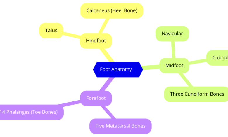
Parts of the foot: The human foot is a remarkable structure, comprising 26 bones, 33 joints, and over 100 muscles, tendons, and ligaments. It is traditionally divided into three regions: the hindfoot, midfoot, and forefoot. Each region plays a crucial role in providing support, balance, and mobility.
Hindfoot
The hindfoot forms the back portion of the foot and includes two primary bones: the talus and the calcaneus. The talus is the ankle bone that links the foot to the lower ends of the tibia and fibula from the ankle joint. The goal of this joint is to regularly move in an up and down direction, and it is used in walking and running. Below the talus is the calcaneus, also referred to as the heel, and is the largest bone in the foot. It is used to establish the stand and support the body weight during movements by locomotion.
The talocalcaneal joint is located at the posterosuperior aspect of the foot; it moves only in a side-to-side direction; inversion is the medial movement, while eversion is the lateral movement. These movements are necessary for responding to emergent surfaces and keeping steadiness. Also, the Achilles tendon connects to the calcaneus and is involved in the lifting of the heel during walking or running.
In summary, the hindfoot’s structure and function are fundamental for absorbing shock, providing stability, and enabling a range of movements necessary for daily activities.
1. Talus
The talus is the smaller one of the 26 bones located in the human foot and is of vital significance. It links your foot to those two lower leg bones called the tibia and fibula in order to create your ankle. This joint lets your foot slide up and down, so you are able to walk, run, and even climb up the stairs. The talus has no muscles but is an important structure responsible for weight transfer from your leg to the foot. It is strong and assists in preventing any vibrations during the transfer of movements from one position to another.
2. Calcaneus
The calcaneus is the heel bone and is also the largest bone of your foot. Lie under the talus, which takes most of your body weight when you are standing, walking, or even running. The Achilles tendon is the tendon that connects to the calcaneus to lift your heel when you are in motion. It is like a pillar or the ground that you stand on to perform day-to-day activities on your feet.

Midfoot
The midfoot consists of five irregularly shaped bones: navicular, cuboid, and three cuneiform bones – medial cuneiform, intermediate cuneiform, and lateral cuneiform. These bones create the shape of the sole of the foot, the structures which, in effect, support and mold and absorb the stress of the body weight when bearing load. They also help carry body weight across the foot and help to adapt to the ground during movement.
Four small ligaments and the plantar fascia, a wide band of fibrous tissue, help to connect the midfoot. Plantar fascia is a structure that runs from the heel to the metatarsal head and gives both stability to the arch and enables the foot to ‘flatten out’ and ‘Store energy’ during the stance phase of walking or running. Muscles from lower leg areas, for example, muscles of the tibialis posterior, help support arches and movement in midfoot bones.
The midfoot’s design allows it to function as a rigid lever during push-off phases of gait and as a flexible structure when accommodating uneven terrain. This adaptability is essential for efficient locomotion and balance.
1. Cuboid
The cuboid is a small cube-like bone that lies on the outer aspect of your midfoot. Immature proximally with the calcaneal surface and distally with the metatarsals and proximal phalanges. Cuboid also plays a role in providing support to the longitudinal arch of the foot and allows the foot to stand and to ‘slide’ when we are walking or running.
2. Navicular
The navicular is a round or semi circular-shaped bone lying in the medial midsection of your foot a little distal to the ankle bone known as the talus. It attaches itself to the cuneiform bones and aids in maintaining the arch of the foot as strong and as supple as possible.
3. Medial, Intermediate, and Lateral Cuneiforms
These three small bones lay parallel to one another in the midsection of your foot. The medial cuneiform is proximal to the big toe; the intermediate cuneiform is in the middle, and the lateral cuneiform is distal to the outer side. In combination, they assist in shaping the foot and its movement and balance.
Forefoot
The forefoot consists of metatarsals and phalanges. The metatarsal bone, number five, it articulates with the proximal phalanges of the toes. The first toe, called the hallux, has two bones and each of the other four toes has three bones. This naturally allows for a great variety of movements, including the basic movements called flexion and extension, that are useful for balance and push-off during walking and running.
It is apparent that much of the body weight is shifting onto the forefoot, particularly throughout the push-off phase of gait activity. Particular importance in this process is the metatarsophalangeal joints, which are located between the metatarsals and phalanges and help the toes move well and push off the ground.
The forefoot is designed to swivel in response to the ground to support the lower leg and balance. Bones and soft tissue distribution in this area provide the foot flexibility during movements, hence improving locomotion.
Five Metatarsal Bones
Metatarsal bones are the five elongated bones of the forepart of the foot. It joins your toes to the mid part of the foot area of your foot without much fuss. These bones are in the order of five, from the first toe to the fifth toe. Metatarsals are the long bones in your foot that hold weight when you stand, as well as offer support and stability in walking, running, or jumping. They are also relevant when it comes to extending your body towards the forward motion during movement.
14 Phalanges
They are those bones that form the structure of the toes, which are the type used by doctors to refer to as the fetal bones. Every toe consists of three phalanges, while the first toe has two phalanges. The phalanges cause the movement and flexibility of your toes, enabling you to balance and possess solid ground when walking or standing. They also cushion your body and ensure you’re steady, especially during the actions and movements. Both metatarsals and phalanges are important to support the movement of the feet.


